Contact: Janese Heavin, heavinj@missouri.edu
Photos by Abbie Lankitus, Nicholas Benner and Nicholas Andrusisian
August 19, 2025
There’s wonder in research — not only in the breakthroughs that change lives but in the breathtaking magic of discovery itself. From the delicate structure of a molecule to the symmetrical geometry of cells, science reveals a world as visually stunning as it is intellectually profound.
At the University of Missouri, that beauty is on display at the Christopher S. Bond Life Sciences Center as part of the Joy of Discovery sculpture. For nearly 20 years, the DNA-shaped installation has helped celebrate the awe-inspiring intersection of art and science, showcasing images captured in labs across campus.
Now, Joy of Discovery has been updated with a new collection of scientific imagery. Selected from more than 40 campus-wide submissions, these images open a window into the hidden processes that happen just below the surface.
Created by artist Kenneth F. vonRoenn Jr. with funding from Alvin E. and Mary Agnes McQuinn, Joy of Discovery was dedicated in 2007.
Scroll down to see scenes from the installation and to learn more about the science behind the art.
What am I looking at?
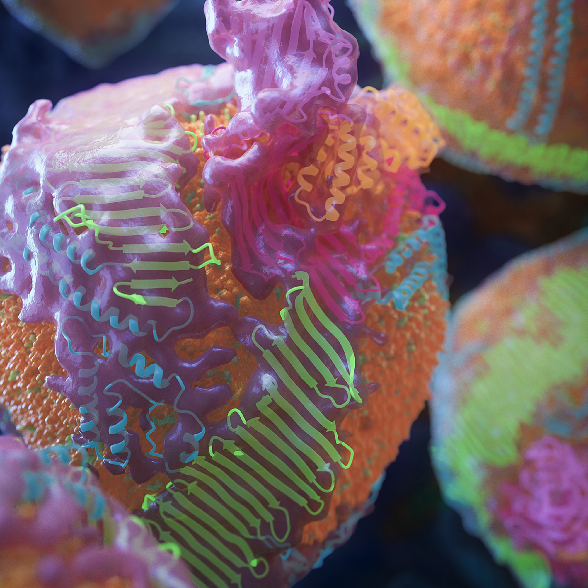
The first full structure of human apoB100 in low-density lipoprotein.
Why does this research matter?
Apo-B100 is a protein that plays a crucial role in carrying cholesterol and other fats through the bloodstream. Understanding it could lead to better ways to assess cardiovascular disease risk and develop targeted treatments.
How was this image created?
Researchers combined cryo-electron microscopy, AlphaFold2 structure prediction and molecular dynamics flexible fitting to build the model — then used a software called MolGraphics using Cinema 4D to create the image. Zachary Berndsen, assistant professor of biochemistry, collected and processed Cryo-EM data from the Titan Krios microscope at the Mizzou Electron Microscopy Core facility. Keith Cassidy, assistant professor of biological physics, performed the molecular dynamics fitting using Hellbender high-performance computing resources.
Where is this work happening?
College of Arts and Science; School of Medicine; Roy Blunt NextGen Precision Health Building
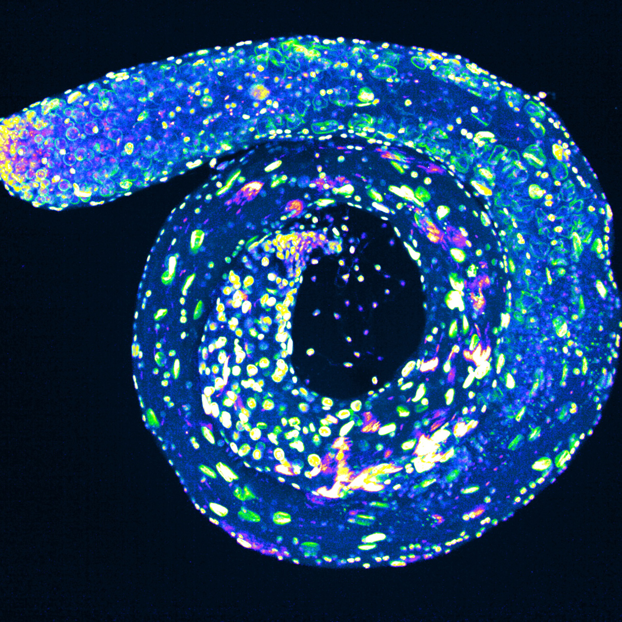
What am I looking at?
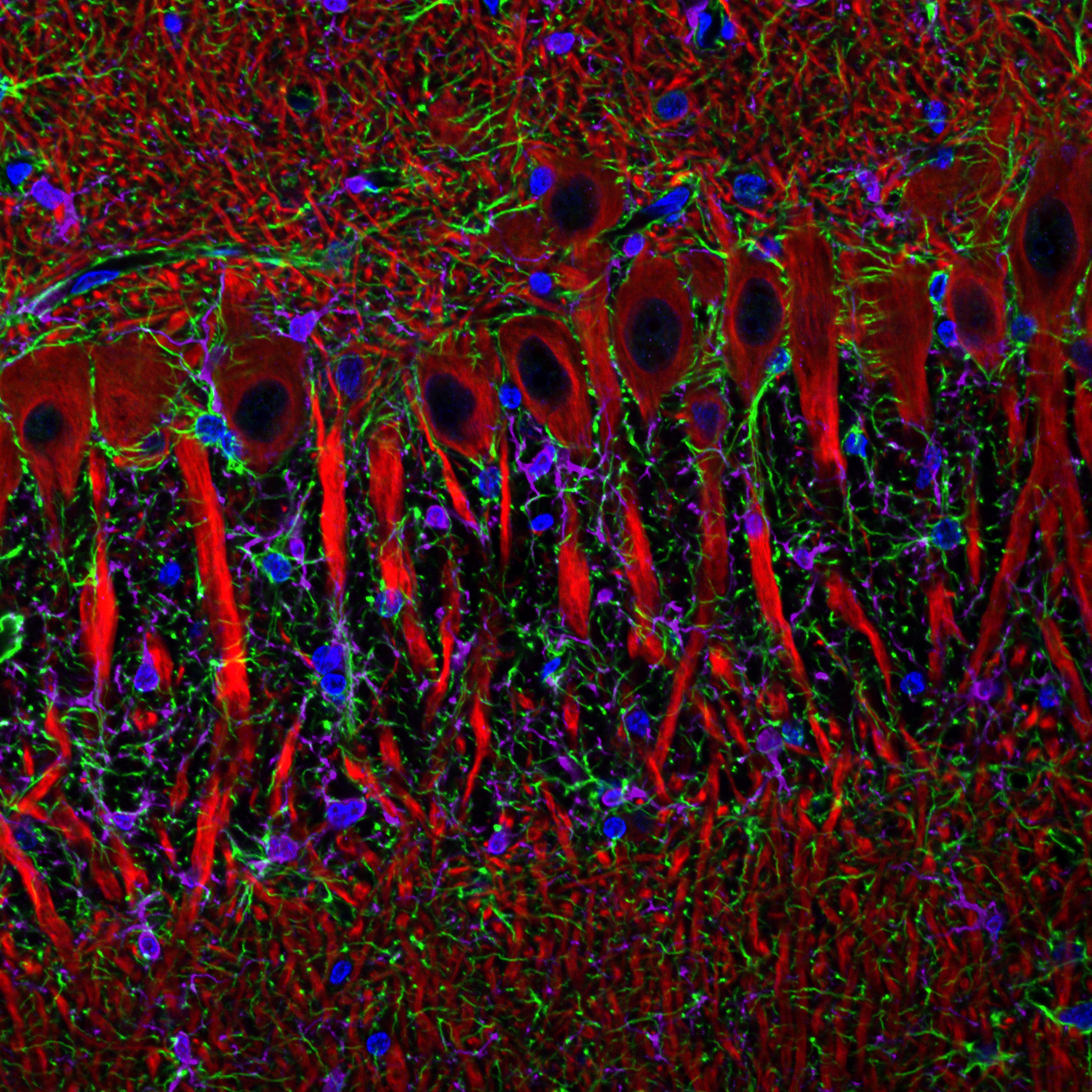
A thin slice of frozen brain tissue — about one-quarter the width of a human hair — from the hippocampus of a cloned pig. The image shows interactions between brain support cells and neurons.
Why does this research matter?
This study helps scientists understand how brain support cells, specifically astrocytes and microglia, interact with neurons. It’s a key step toward developing models that more closely mimic human brain diseases and injuries.
How was this image created?
The frozen tissue was stained with fluorescent markers to label different cell types: astrocytes (green), neurons (red), microglia (purple) and cell nuclei (blue). The image was a collaboration between Katherine Rodriguez-Lukey, research specialist at the Bond Life Science Center’s Advanced Light Microscopy Core, and Kiho Lee, associate professor in the Division of Animal Sciences.
Where is this work happening?
College of Agriculture, Food and Natural Resources, Bond Life Sciences Center
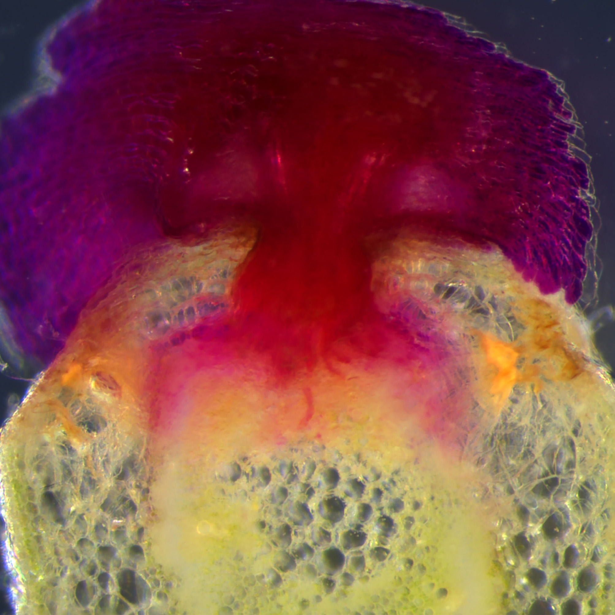
What am I looking at?
A genetically modified parasitic plant producing red pigments parasitizes a tomato host.
Why does this research matter?
Parasitic plants such as Cuscuta, commonly known as dodder, drain water and nutrients from crops such as tomatoes, potatoes and beans, causing agricultural losses. This work allows scientists to genetically modify Cuscuta for the first time, creating new opportunities to study how it attacks its hosts and ways to stop it.
How was this image created?
Soyon Park, assistant professor from the Division of Plant Science and Technology, and her lab members used a genetic marker called the RUBY reporter, which produces red betalain pigments in transformed tissues. This image, taken at the Mizzou Advanced Light Microscopy Core, captures the striking moment when Cuscuta transfers these red pigments to its host, a tomato plant, illustrating the process of parasitism in vivid detail.
Where is this work happening?
College of Agriculture, Food and Natural Resources
What am I looking at?
This image shows the distribution of the hormone abscisic acid (ABA) in a young Arabidopsis thaliana seedling. It highlights the different levels of ABA across different parts of the plant, especially the seed leaves (cotyledons) and the stem-like region below them (hypocotyl). The color intensity in the image represents the concentration of ABA in each area.
Why does this research matter?
The research uses advanced tools known as FRET-based biosensors, which allow scientists to monitor plant hormone activity in real time inside living cells. These sensors help detect early signs of stress, for example, when plants don’t have enough water. The biosensors reveal spatial hormone gradients that provide key insights into signaling pathways, improving phenotyping resolution and enabling in-depth studies of hormone interactions at cellular and tissue levels.
How was this image created?
Images were acquired using a Leica SP8 confocal microscope. Segmentation and emission ratio analysis were performed in a platform called ImageJ/FRETENATOR. The image was created by Norma Castro Guerrero, research scientist in the lab of David Mendoza Cozatl, professor in the Plant Science and Technology division and at Bond Life Sciences Center.
Where is this work happening?
College of Agriculture, Food and Natural Resources, Bond Life Sciences Center
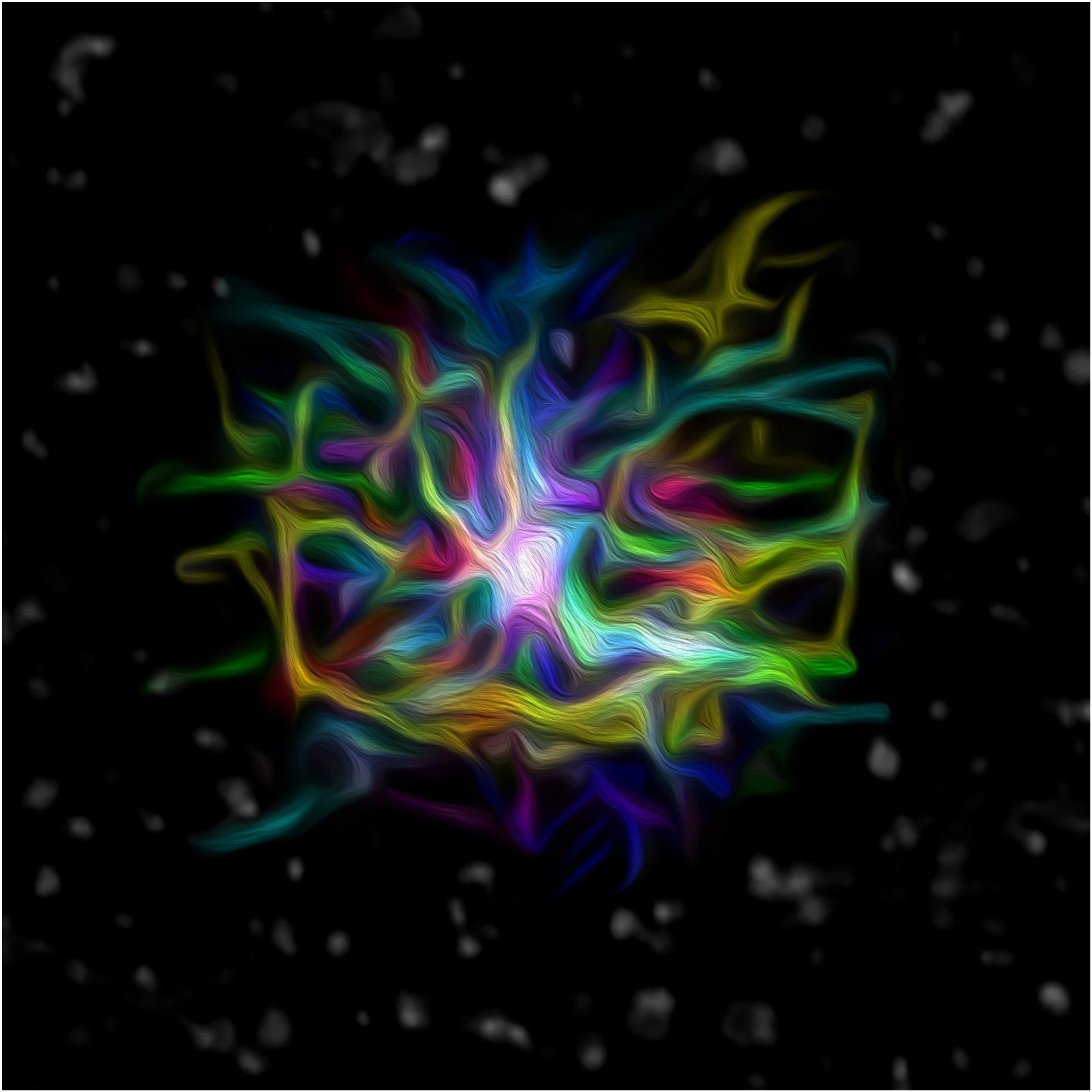
What am I looking at?
A heart muscle cell with glowing star-like shapes made of microtubules and ion channels. The bright white centers represent active TRP ion channels, and the surrounding “stars” are cardiac lysosomes.
Why does this research matter?
TRP ion channels are proteins that, when overactive, can damage heart cells and trigger arrhythmias. By visualizing these channels, researchers can explore new ways to prevent or treat heart rhythm disorders.
How was this image created?
Matthew Hayden, a graduate student, and Timothy Domeier, associate professor of medical pharmacology and physiology, used fluorescent labeling and advanced microscopy to visualize microtubules, TRP channels and lysosomes inside a single heart cell.
Where is this work happening?
School of Medicine
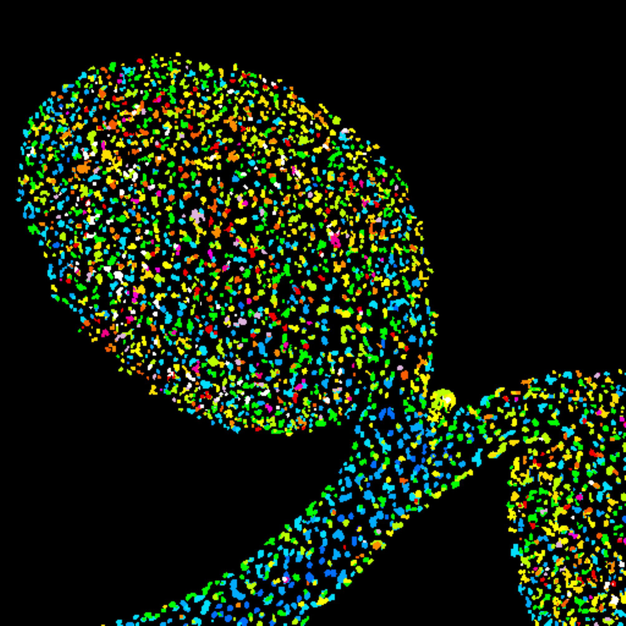
What am I looking at?
The testis of a common fruit fly, or Drosophila, stained to show nuclear membranes and DNA.
Why does this research matter?
This research helps scientists understand how cells divide and organize themselves during tissue formation. The team’s recent work revealed that certain isoforms of a protein known as Mud promote male fertility. The research helps understand fertility and development, not just in flies but in humans and other animals as well.
How was the image created?
Tara Finegan, assistant research professor of biological sciences and director of the Mizzou Advanced Light Microscopy Core, captured the image using a type of fluorescence microscopy as z-stack projection, a technique that combines multiple images taken at different depths. The image was captured at the Mizzou Advanced Light Microscopy Core on an Andor Dragonfly 602 spinning disk confocal microscope.
Where is this work happening?
College of Arts and Science, Bond Life Sciences Center
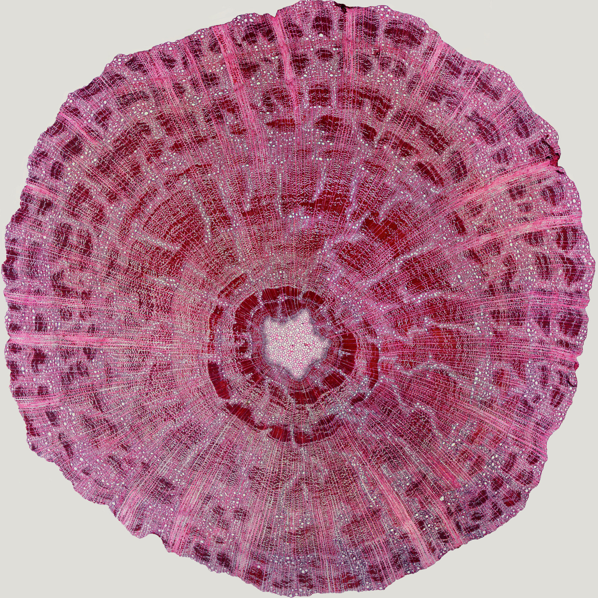
What am I looking at?
A microscopic cross-section of a 20-year-old white oak seedling stem collected from a forest stand under uneven-aged management at the Missouri Ozark Forest Ecosystem Project in southeastern Missouri.
Why does this research matter?
This work allows scientists to understand white oak tree regeneration and growth dynamics— especially by resprouting — and their implications for uneven-aged forest management. These insights can help guide efforts to manage forests and protect our oak-dominated ecosystems in the Ozark Highlands and elsewhere across the eastern U.S.
How was this image created?
Ryan Gross, a graduate research assistant, and Michael Stambaugh, associate professor of forest ecology, prepared the image at Mizzou’s Center for Tree-Ring Science where, for this project, they’ve merged more recent Quantitative Wood Anatomy (QWA) methods with traditional Dendrochronological, or tree-ring dating, techniques.
Where is this work happening?
College of Agriculture, Food and Natural Resources
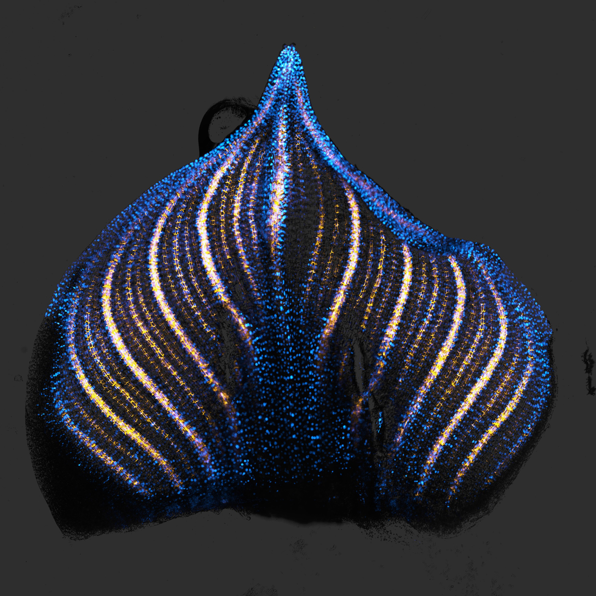
What am I looking at?
A developing maize leaf with colors showing where two key plant hormones are active in the veins.
Why does this research matter?
Understanding how hormones such as auxin and cytokinin guide leaf development helps scientists uncover how plants grow and form complex structures. These insights could help support crop improvement and higher yields.
How was the image created?
Janlo Robil, a PhD student in the lab of Paula McSteen, used confocal microscopy with fluorescent markers. The yellow shows the location of the plant hormone auxin in veins, and the blue color shows the plant growth hormone cytokinin.
Where is this work happening?
College of Arts and Science, Bond Life Sciences Center
"I am honored to have my work included in the Joy of Discovery sculpture. Although my research lab is not located within Bond, I visit the building frequently to meet with colleagues, use the core facilities, and participate in seminars. As the centerpiece of the building, I can’t help but admire the sculpture every time I am in Bond. There is so much amazing science being done at Mizzou, and it is great to see it celebrated in such a beautiful way." -Zach Berndsen