Published on Show Me Mizzou Dec. 17, 2025
Story by Marcus Wilkins, BA ’03
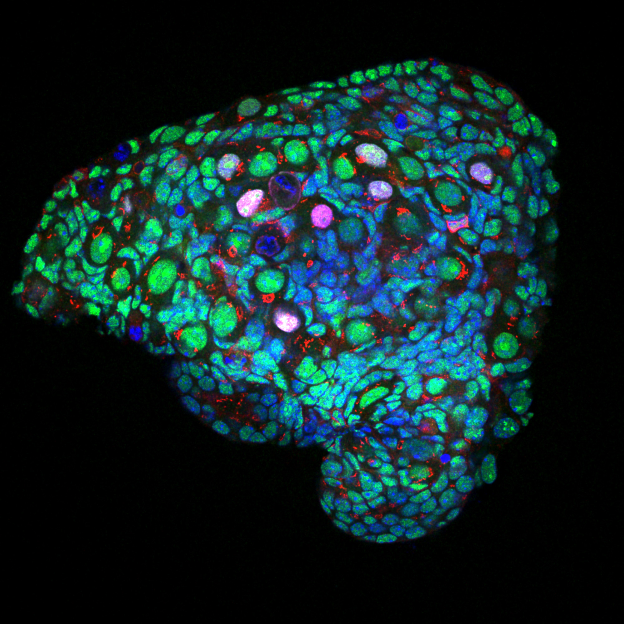
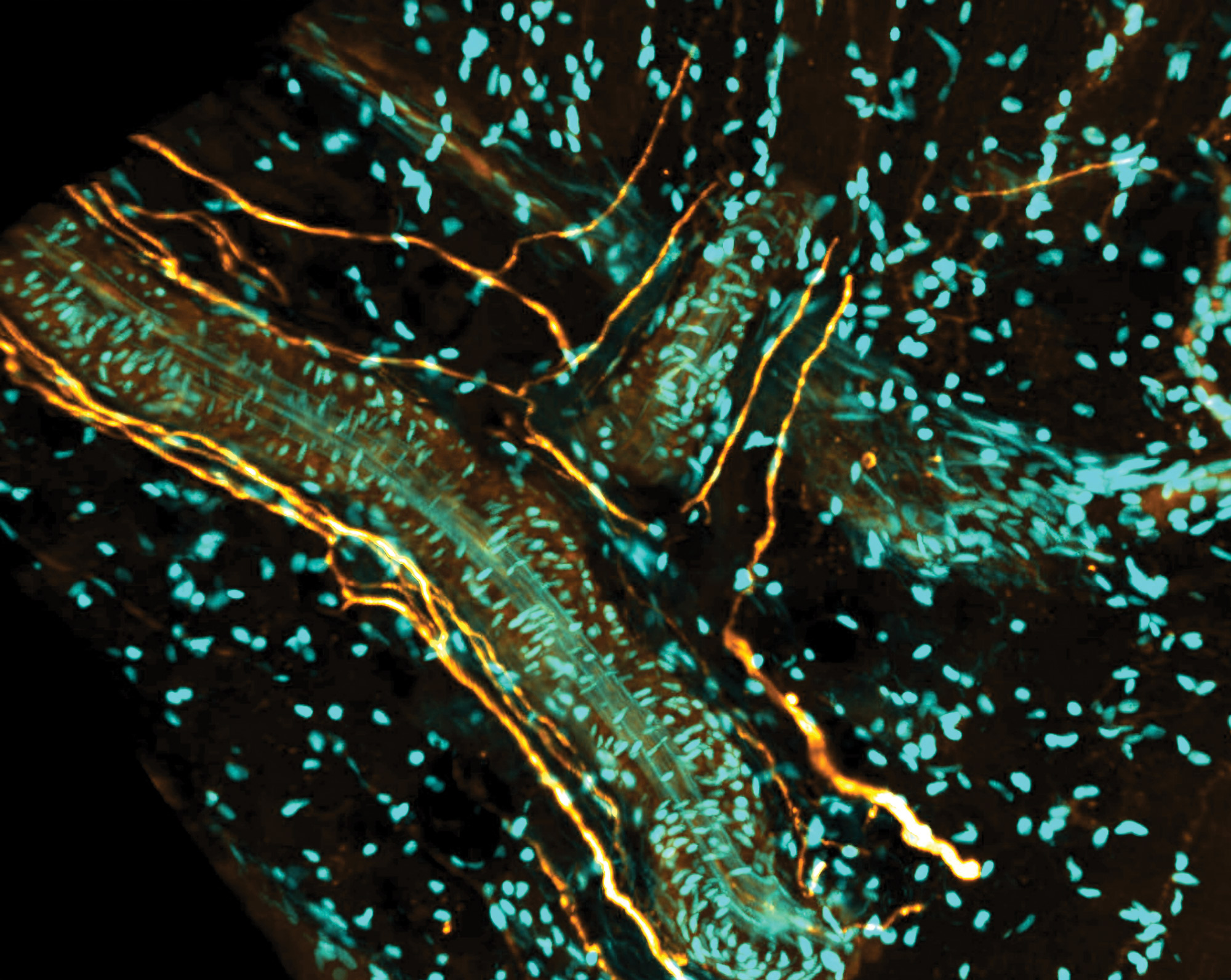
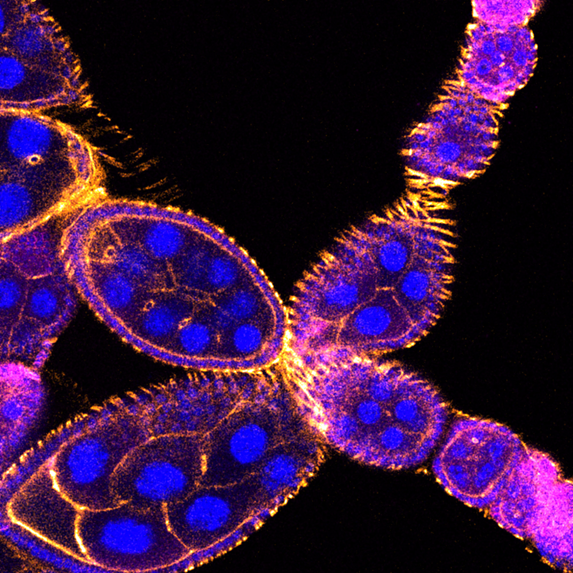
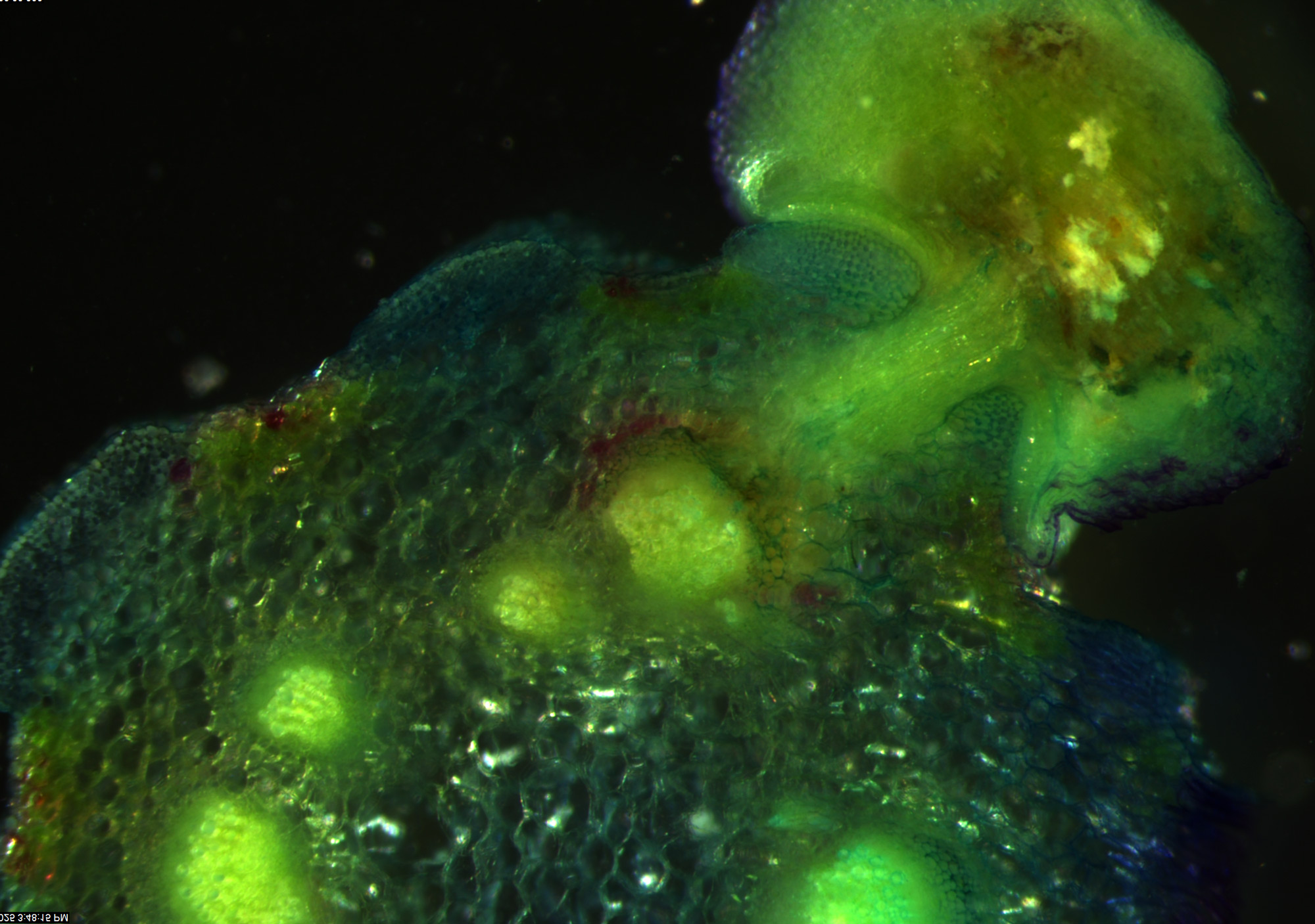
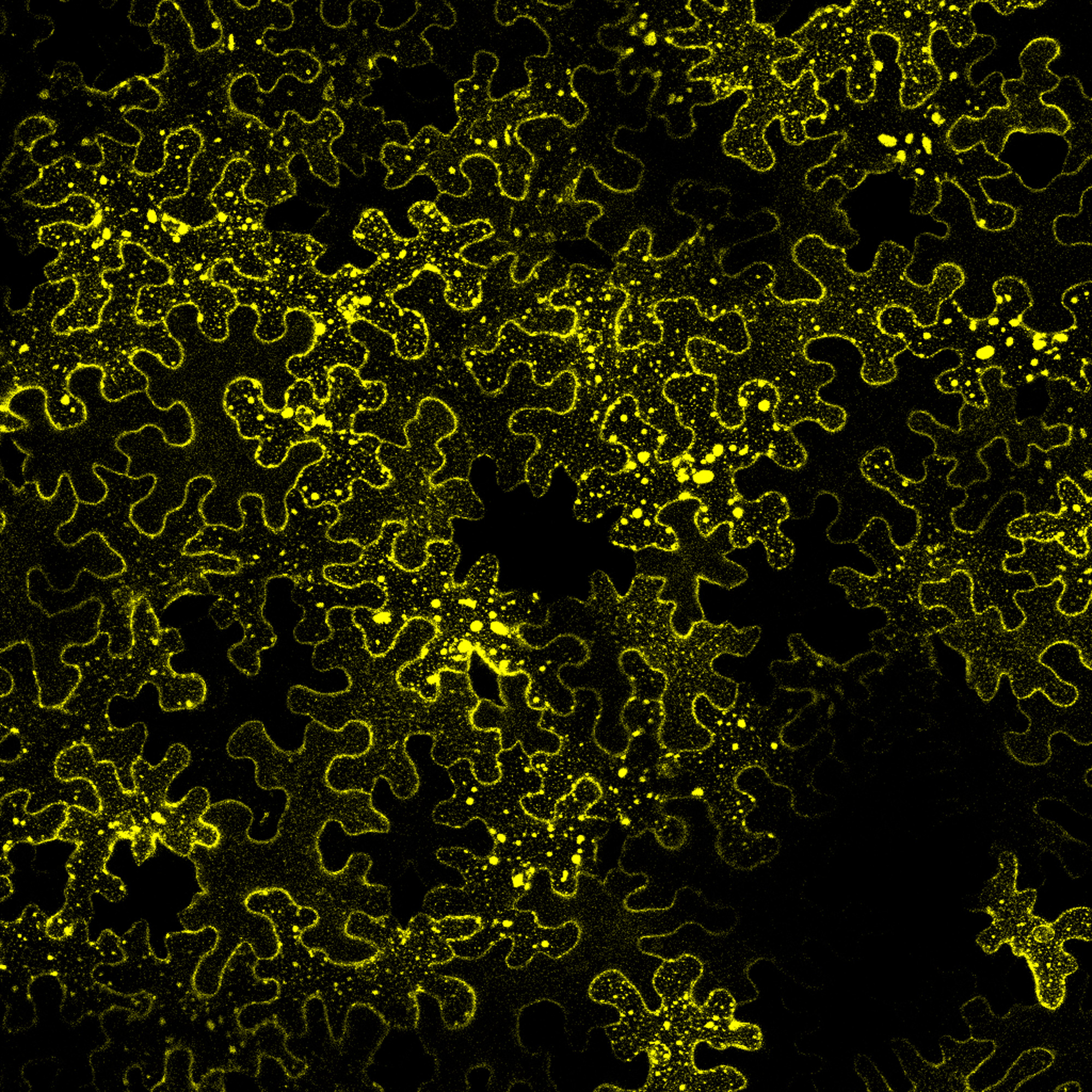
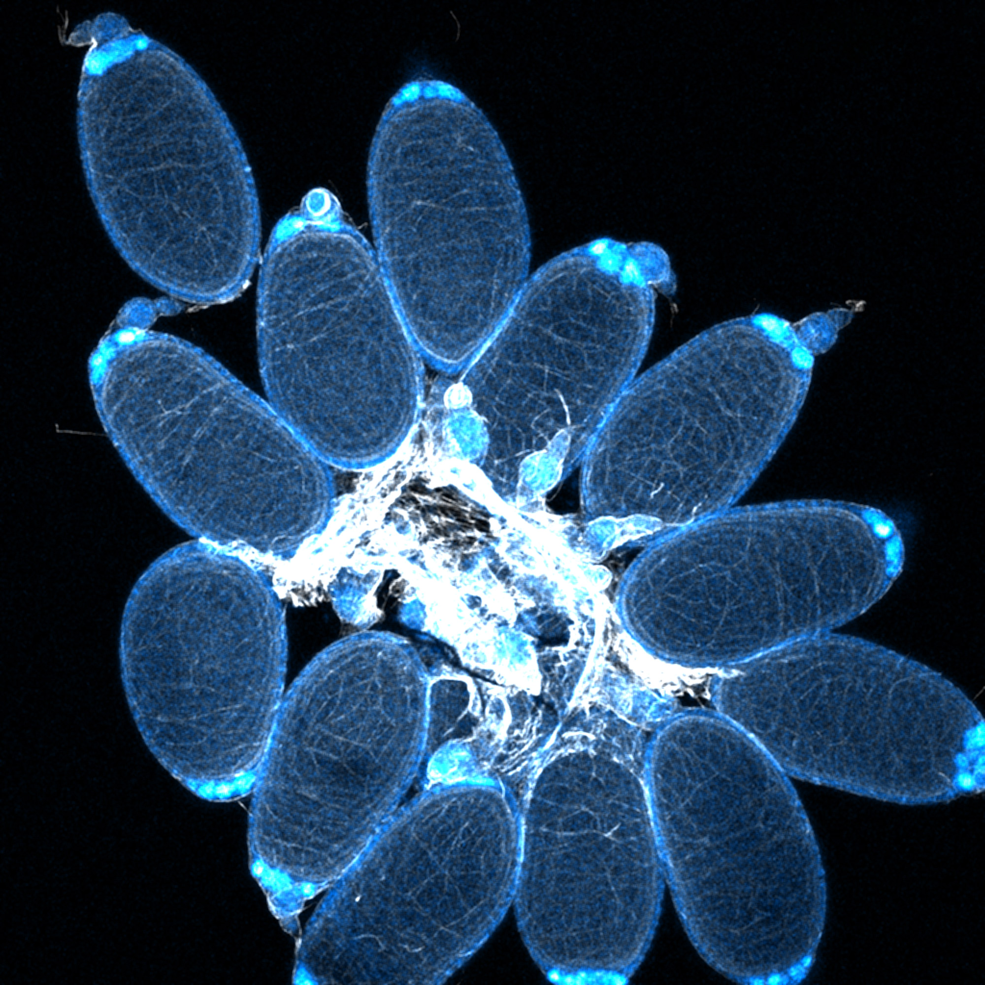
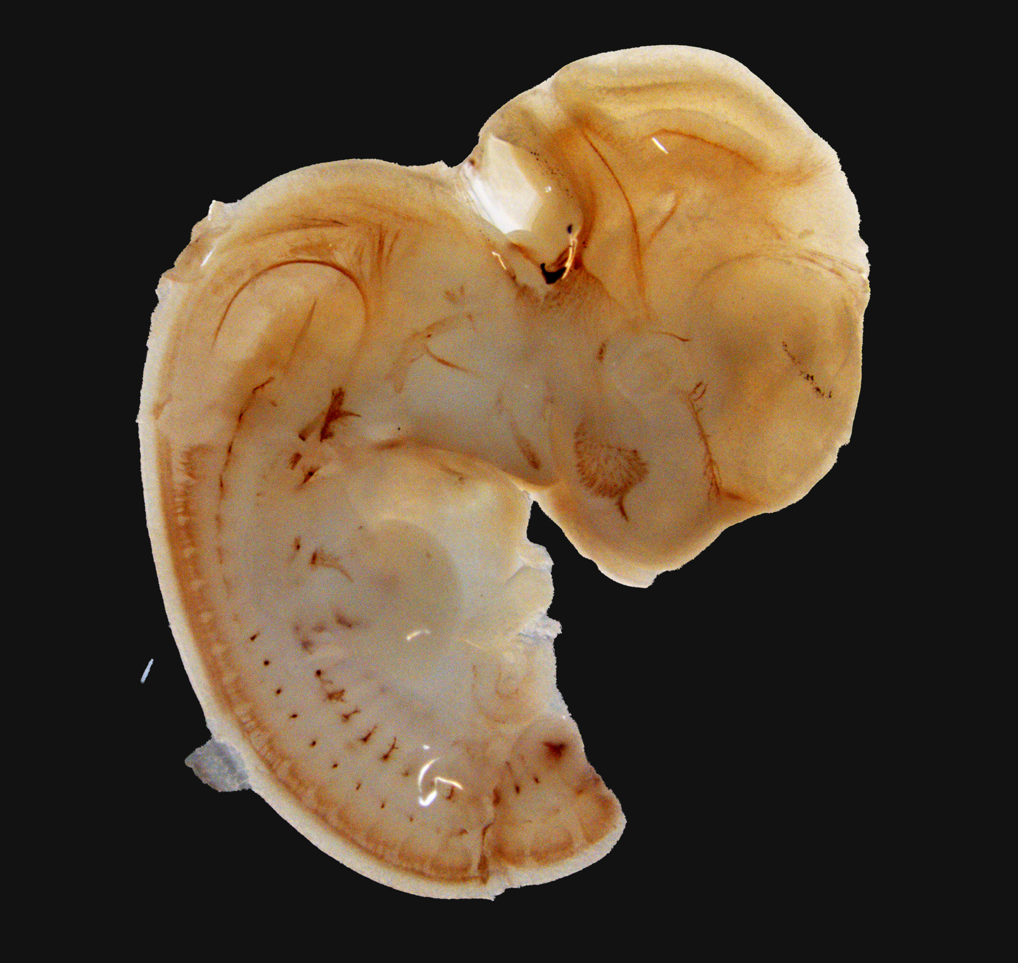
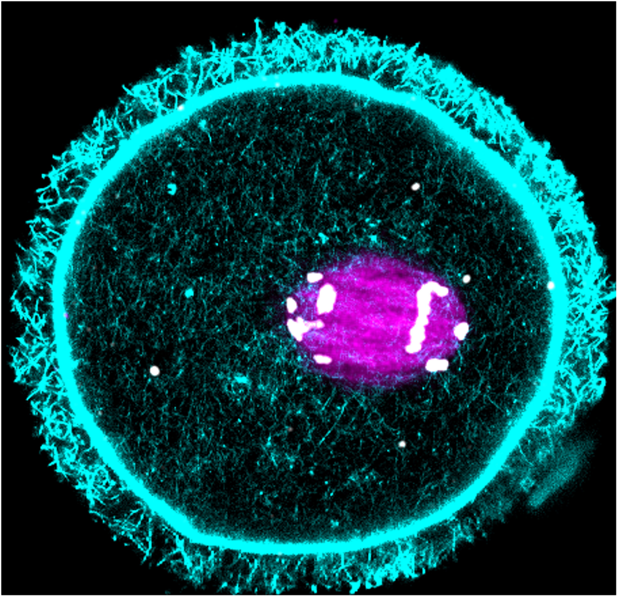
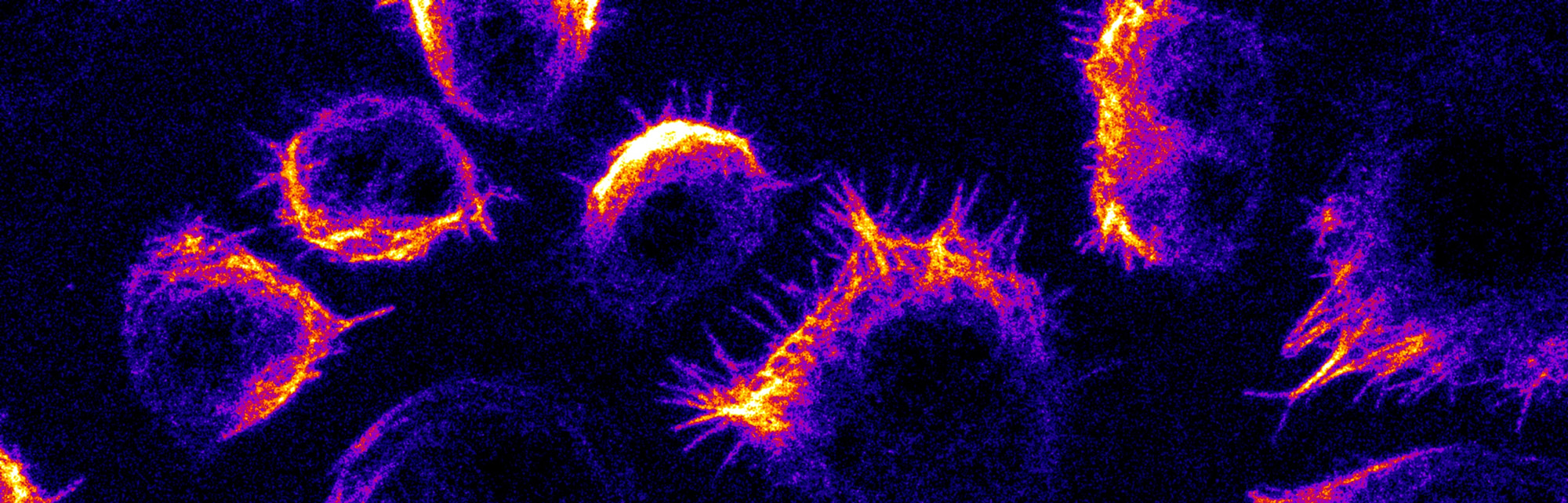
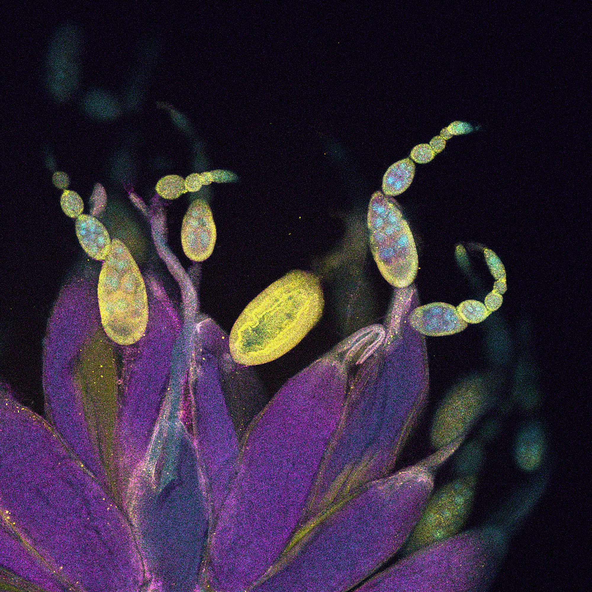
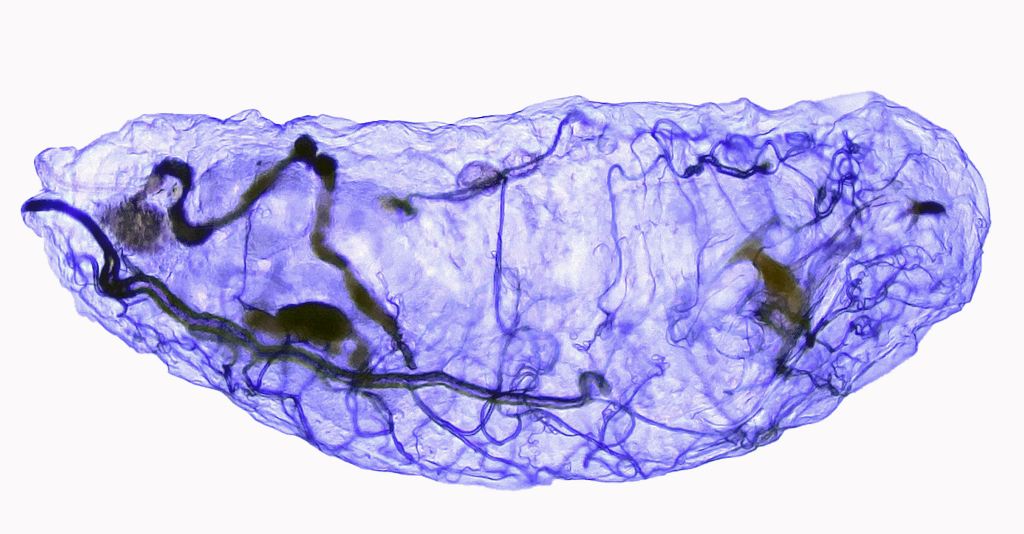
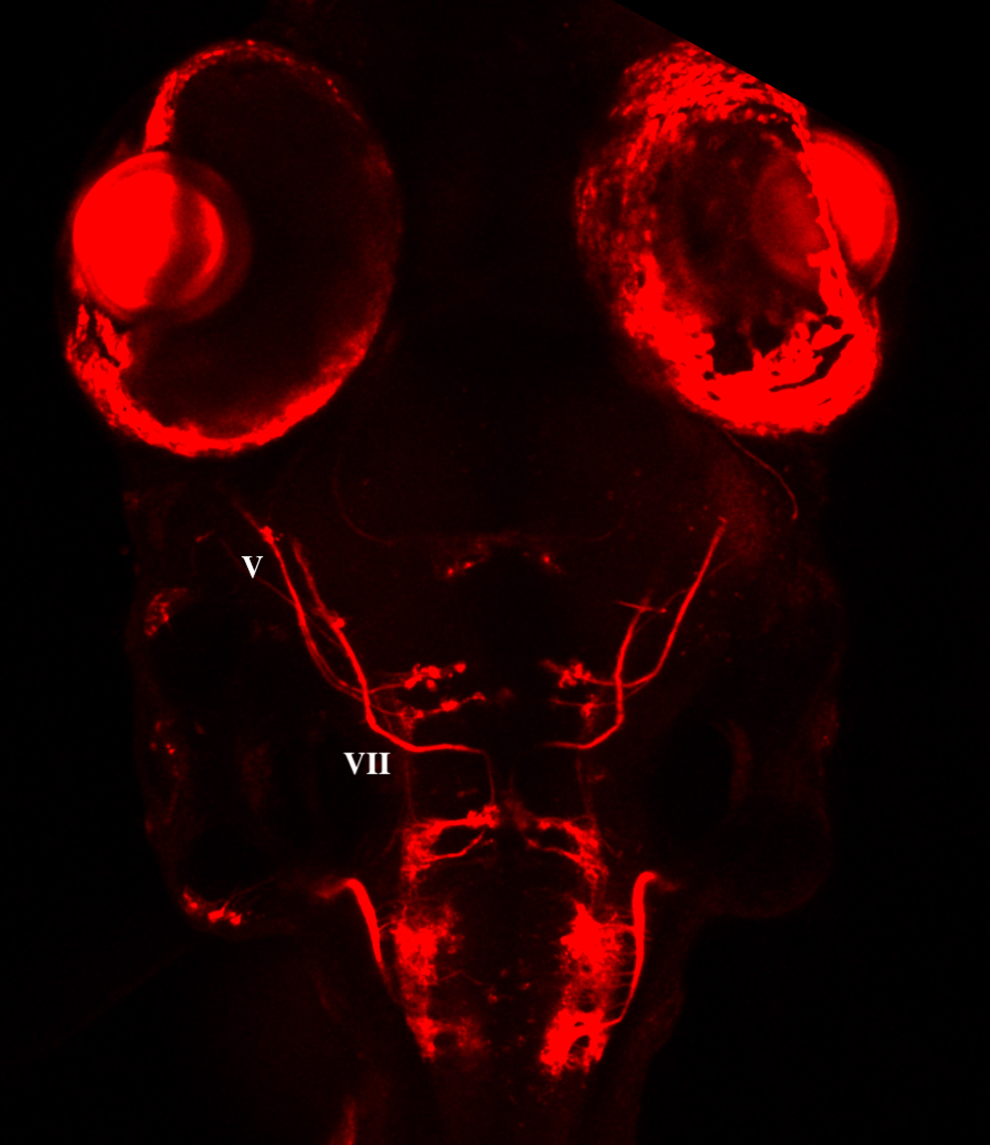
Kaleidoscopic, multifaceted meteors of stained glass. Disembodied, violet-tinted and veinous pods. Luminescent, fluorescent, opalescent bits of ethereal debris. The images created at the University of Missouri’s Advanced Light Microscopy Core (ALMC) reveal a menagerie of alien forms within a planetarium of infinitesimal wonder. The surreal perspectives often feature curiously mundane sources: mosquito ovaries, cabbage white butterfly eggs, zebrafish larvae or the cellular scaffolding that supports beet stems. The mysteries revealed, though, might one day guide research advances in cancer care, orthopedic implant design and food production.
Captured using a fleet of expertly maintained state-of-the-art microscopes housed in Mizzou’s Christopher S. Bond Life Sciences Center, ALMC photos so often are breathtakingly beautiful that the facility conducts an annual contest to celebrate them.
“The first time I visited the ALMC, I was blown away with the instrumentation and the quality of the imaging here,” says Tara Finegan, ALMC director and a Cambridge University-educated researcher. “Collaboration is my favorite part of research, and one reason I am excited to be in this position is that it allows me to work with world-class researchers from across the university.” Finegan was hired as part of MizzouForward, the 10-year, $1.5 billion initiative to strengthen faculty, upgrade infrastructure and enhance student success.
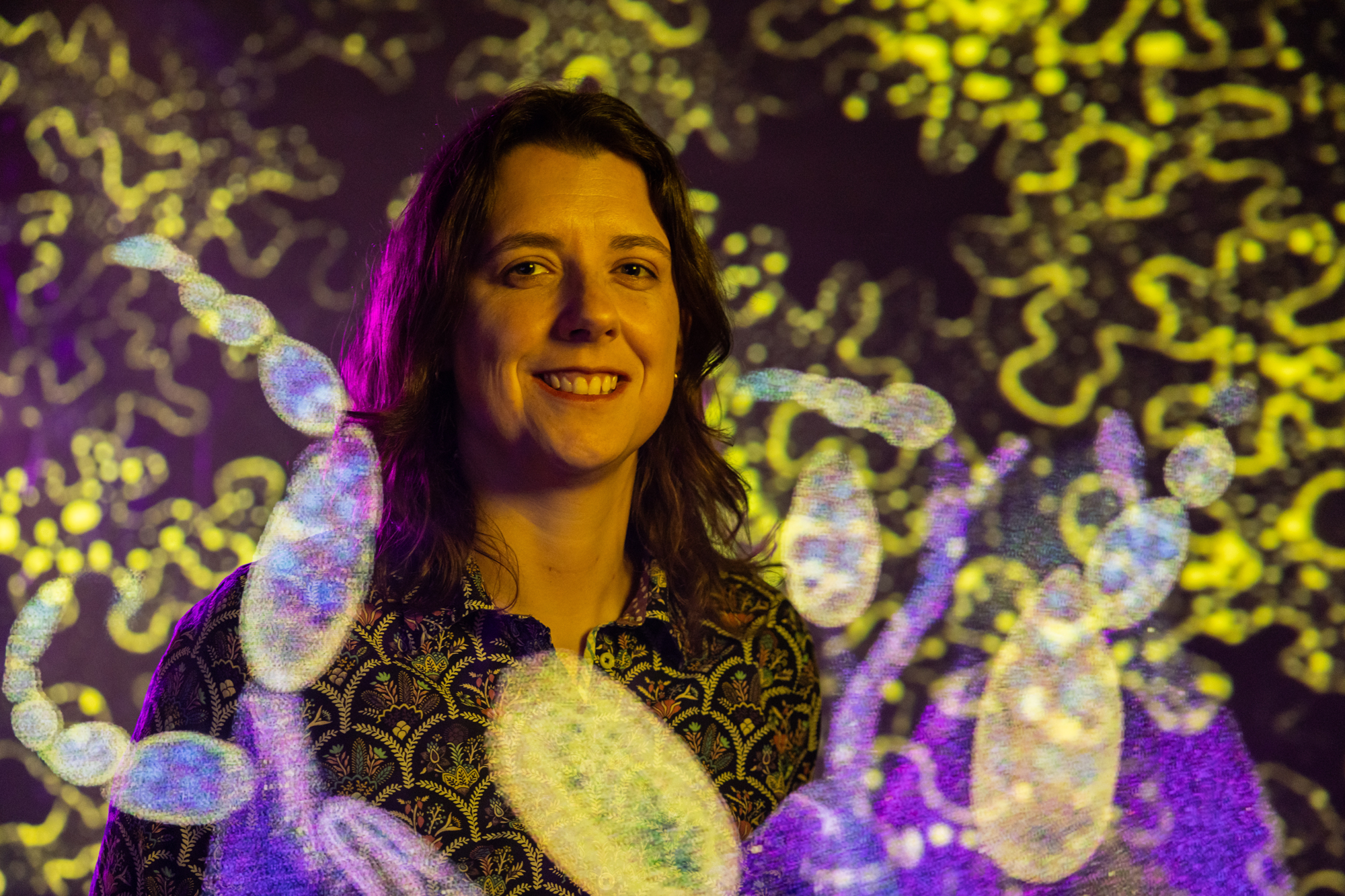
Tara Finegan joined ALMC as its director in 2023, bringing Cambridge training and a passion for collaboration as part of MizzouForward’s investment in research excellence.
The ALMC, one of 17 Advanced Technology Core Facilities supported by the university’s Division of Research, Innovation & Impact, sits within a network that includes the Bioinformatics and Analytics Core, the Genomics Technology Core, the Metabolomics Center and many more across campus. The ALMC houses 11 light microscopes (as opposed to electron microscopes, which use beams of electrons to view much smaller objects) along with an array of related equipment.
Researchers capture ALMC images using one of three types of microscopes:
• Widefield microscopes are the classic variety. They shine light through the whole sample at once to capture an image but tend to blur above and below the focal point.
• Confocal microscopes use lasers and a small pinhole to focus on just one thin layer of a sample at a time, which cuts blur from other layers and provides detailed 3D images.
• Stereomicroscopes are used to view larger, solid objects, such as insects or plants, in 3D and at a lower magnification. They also allow researchers to manipulate or dissect specimens while viewing.
Among the core’s services, staff train visiting researchers to use the technology before independently embarking on their explorations.
“The ALMC is an invaluable resource to my lab’s research,” says David Braun, professor of biological sciences and plant sciences. “We use their state-of-the-art microscopes to image proteins and cell structures to investigate their functions, and the staff are incredibly knowledgeable, highly experienced and always helpful.”
For example, staff might assist with preparation by using a microtome, a precise slicing instrument for making thin samples of tissues. Staff also help with quantitative image analysis — counting and measuring things — using AI-assisted software to extract data.
Lei Lei, associate professor of obstetrics, has used the core to research cancer growth. “Our research examines how egg cells undergo cell death during cancer treatment, and the ALMC enables us to look deep inside mouse ovarian tissues to precisely examine the molecular and morphological features of different components,” she says. “The knowledge helps in developing medical interventions to preserve fertility for young female cancer patients.”
The core’s suite of rooms housing its microscopes is named after Thomas E. Phillips, the core’s founding director and Curators’ Distinguished Teaching Professor Emeritus. The core has seen increased usage since 2021 thanks in part to MizzouForward bringing an influx of biomedical researchers. Work completed at the facility has resulted in 22 scientific publications during fiscal year 2024–25. The ALMC also provides hands-on research experience for undergraduates connected through the Office of Undergraduate Research.
Each frame captured at the core is a constellation of color and code, a reminder that life’s deepest mysteries begin in miniature. “At the ALMC, we make the invisible visible,” Finegan says. “From single molecules to whole tissue and organisms, our microscopes, and the researchers who operate them, reveal how living systems are built and how they change — insights that drive progress in health, agriculture and biotechnology.”
To read more articles like this, become a Mizzou Alumni Association member and receive MIZZOU magazine in your mailbox. Click here to join.