Oct. 24, 2024
Coughing was just a fact of life for Hannah Gryder.
For as long as her grandmother, Tina Wood, can remember, the rasping noise and chest spasms would disrupt Hannah’s sleep. She was diagnosed with asthma at five and got sick with pneumonia and bronchitis several times.
In their search for answers, they went to allergy specialists and tried allergy shots. Hannah, now 12, had her tonsils and adenoids removed, hoping that would help. Wood even completely redecorated their home in Pleasant Hope, Missouri, outside of Springfield, in case she was allergic to something in the house.
But Hannah’s cough persisted as she moved through middle school. She started seeing a pediatric pulmonologist in Springfield, who diagnosed her with a chronic lung condition called bronchiectasis. Hannah’s airway was thicker than normal because of extra mucus that made infections more common.
She was referred to MU Health Care’s Children’s Specialty Center, where testing from the cystic fibrosis team, including Rebekah Nevel, confirmed that Hannah has cystic fibrosis.
Cystic fibrosis, or CF, is a genetic condition that affects the body’s ability to regulate chloride salt in cells. Salt channels are how the body changes the amount of water in cells.
Less water means thicker, stickier mucus, which causes damage to lung tissue and makes airway infections more likely. CF can also lead to liver, gastrointestinal, pancreas and endocrine problems.
“Cystic fibrosis takes a team to care for it,” Nevel said. “As one of four CF centers in Missouri, we have everything in place to help pediatric patients, like Hannah, and adult patients prevent lung disease progression and live their lives to the fullest.”
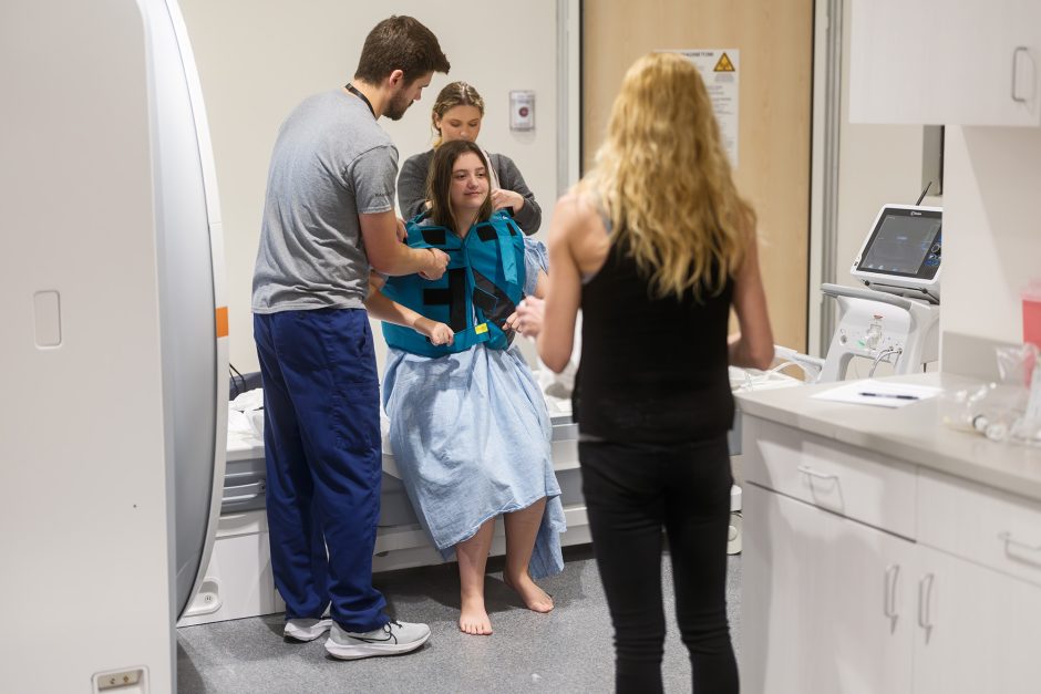
But Nevel, and everyone else involved in Hannah’s care, has access to something most CF centers do not. Steps away from her clinic, Nevel can walk into the Roy Blunt NextGen Precision Health building to get the clearest images possible of her patients’ lung function using an MRI machine and xenon gas.
Before this technology was developed by MU Health Care and School of Medicine researchers and radiologists Talissa Altes and Robby Thomen, pulmonologists were unable to use MRI to see a diagnostic image of lung function, relying on useful but imperfect tools like CT scans, spirometry and plethysmography.
This FDA-approved clinical scan can show doctors at MU Health Care how healthy a person’s lung tissue is, and how efficiently oxygen and carbon dioxide are exchanged in the lungs. Hannah was one of the first patients at MU Health Care to receive a xenon MRI scan for clinical care.
“Hannah’s results helped Dr. Nevel to suggest some medications that would help Hannah’s treatment because she could see more clearly the things that were going on in her lungs,” Wood said.
Hannah and her family have made a few trips to Columbia for the xenon MRI scans. But most of her ongoing cystic fibrosis care happens at MU Health Care’s cystic fibrosis outreach clinic in Springfield, much closer to home.
Read more from MU Health Care


