Bridging the gap from an initial idea to the commercial market takes years of research and development in the lab, a team of dedicated researchers and, of course, funding. That’s where the Missouri Tech Expo comes in.
At the Sept. 19 event on the MU campus, researchers from MU and other University of Missouri System schools will have the chance to woo investors with elevator pitches. They have 10 minutes to present what they’ve developed and explain why the world needs access to it.
Here are three MU research teams working to get their innovations to market:
Screening for autism

Dr. Gary Yao and Dr. Judith Miles could potentially change the way physicians screen very young children for autism spectrum disorders.
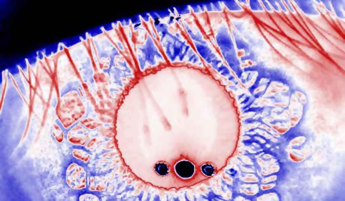
With the expertise of Miles, professor emerita for child-health and genetics practicing at The Thompson Center, Yao, a professor of bioengineering, and Randima Dinalankara, a graduate student in bioengineering, have developed a device that measures pupillary response as an indicator of autism and other neurodevelopmental disorders.
The device measures the pupil’s response to light, how long it takes for the pupil to react and how much the pupil constricts.
“In order to accurately measure the pupil’s response, we have to take a high-resolution image of the pupil at high speed,” Yao says.
Taking a clear image can be difficult with wiggly children younger than 5 or 6 years old. This device, however, tracks the child’s movement and captures images without requiring the child to remain still. Children can even sit freely on their parents’ laps watching movies, and the image will still be clear.
About one in every 100 children has an autism spectrum disorder. Early detection is key because it allows for early treatment. Currently the most common form of screening for autism is behavioral observation.
“This device would be a much more exact, valid screening process,” Miles says.
The goal is to get the device into the offices of pediatricians who can incorporate it into regular well-child checkups.
“We think it’s very exciting, and we don’t want to keep the light under the basket,” Miles says. “If you just use this in your lab and say ‘I’m not going to share it with the rest of the world,’ you really haven’t done your due diligence to patients, their families, as well as science.”
Diagnostic kit for anticancer treatment

Dr. Gerald Arthur, Dr. Raghuraman Kannan and graduate student Chuck Caldwell have developed a diagnostic kit to help oncologists more effectively treat cancer patients.
Using nanotechnology, the kit detects overexpressed biomarkers (biological indicators of some state or condition) present in human tumor tissue.
“Based on the concentration of biomarkers, we can say whether a particular targeted drug is going to work for a patient or not,” says Kannan, associate professor of radiology and biological engineering. “We can prescribe the targeted medicine to suit the patient.”
Many targeted anticancer drugs are highly expensive, running anywhere from $30,000 to $80,000. Not all patients respond to these drugs, however.
“We can cut down the number of ineffective drug treatments by properly classifying the tumors,” says Arthur, research assistant professor for the Department of Pathology and Anatomical Sciences.
“We believe we can better identify those patients who will respond and therefore get the benefit of the drug, and we can also identify the patients that aren’t likely to respond and therefore prevent any side effects of the drug,” Arthur says.
Currently, the research is supported by the Wallace H. Coulter Foundation. The team was awarded an additional $100,000 by the foundation in September through The University of Missouri’s Coulter Translational Partnership Program.
The team is in the process of forming a company. The next step in bringing the kit to the commercial market is to get it approved by the Food and Drug Administration.
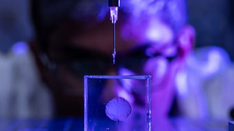
Ultra-stable atomic force microscope
Dr. Gavin King and his team of researchers have designed and built an ultra-stable atomic force microscope (AFM), which uses lasers, optics and computer software to provide a more complete image of nanoparticles in three dimensions.
Traditional atomic force microscopes, or AFMs, capture only one-dimensional information from samples.
King, an assistant professor of physics and biochemistry, says the traditional process is like running your finger over Braille; you only feel the tops of the bumps. Using an ultra-stable AFM, he says, is more like running a finger over your knuckles. You can feel all the troughs and crests of the bumps.
Because the ultra-stable microscope captures that extra information, it provides scientists with better and more complete images of their samples.
“It’s a really non-invasive way to study individual molecules in near-native conditions,” King says. “What separates a biological structure from an inanimate object is the fact that it moves and responds to stimuli. This microscope also allows you study these molecules as they undergo dynamics.”
This feature, King says, will be crucial to biologists.
Potential applications for the microscope include the discovery of new drugs and the study of drug binding, which could lead to better pharmaceuticals.
The next big thing
Expo highlights include talks by entrepreneurs currently heading biotech companies. Han Chen, managing director of the technology incubation firm Kapyon Ventures, gives the morning keynote address. Later, attendees can hear from Karl Kochendorfer, CEO of the clinical-decision support system firm MedSocket, and from Jeremy Paben, vice president of engineering for EyeVerify, which makes mobile-device security technology based on blood vessel patterns in the user's eyes.
Now in its fourth year, the Missouri Tech Expo annually honors MU research teams that have successfully commercialized their research. Past success stories include:
- Organovo: Professor Gabor Forgacs and his research team developed 3-D bioprinting. Their company, Organovo, is now traded on the New York Stock Exchange under the symbol ONVO.
- Beyond Meat: Professor Fu-Hung Hsieh and his team developed a soy-based meat substitute that replicates the taste, texture and appearance of chicken. This technology is licensed to Savage River Farms Inc. and commercialized under the brand name Beyond Meat. The product is manufactured in Columbia, Mo., and is available in grocery stores nationwide.
- EternoGen: MU Biodesign and Innovation Program fellows and Professor Sheila Grant and her research team discovered that gold nanoparticles extend the life and stability of collagen. After successfully attracting more than $1.5 million in investments, their company, EternoGen, announced its plans to enter the commercialization phase.